Introduction

Introduction

Cancer is a common yet devastating disease caused by mutation of cellular DNA and loss of cell cycle regulation (Campbell and Reece). Such mutation, called transformation, occurs as a result of exposure to mutagenic factors such as radiation and UV rays or with no apparent cause (Campbell and Reece). Cancer is expected to claim 13 million lives annually by 2030, and 21.7 million individuals are expected to be diagnosed annually by that year (“Global Cancer Facts & Figures”). Cancerous cells multiply rapidly, forming tumors, or masses of cancerous cells, and may metastasize, or migrate to other parts of the body, through the blood or lymphatic system, which poses an even greater danger (Campbell & Reece, 2007). Cancerous cells disrupt bodily activities as they consume resources as well as disrupt tissue structure and function (Campbell & Reece, 2007).

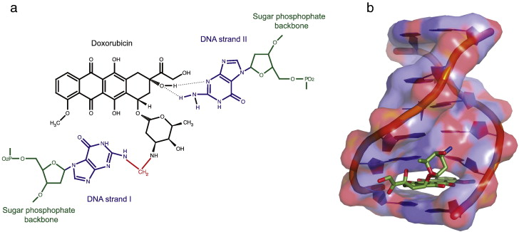
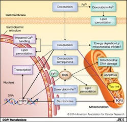
Any drug or treatment that can destroy cancerous cells, therefore, is of prime importance. Tumors of appropriate size, location, and type may be surgically removed. After surgery, swelling, drainage, bruising, or infection can result at the site of surgical treatment. Pain is common, and appetite loss and fatigue often result (Cancer.net Editorial Staff, 2016). Targeted radiation is used to destroy cancerous cells as their mutated genomes are more susceptible to further damage. However, radiation therapy can still damage healthy cells exposed to radiation near the irradiated site (Cancer.net Editorial Staff, 2017). These common treatments, however, fail to fight cancer outside a specific location; instead, chemotherapy treatments use chemicals that circulate throughout the body and destroy cancerous cells as well as some healthy cells (“Chemotherapy to Treat Cancer”). This often results in hair loss and blood disorders, since chemotherapeutic drugs damage hair follicles and inhibit the production of red blood cells in bone marrow (“Chemotherapy to Treat Cancer”). As with other treatments, pain and nausea can result. These chemicals may include those that inhibit specific components of cell division or the cell cycle or may cause cell death (Yang, et al). Doxorubicin, a specific cancer drug part of the anthracycline class of anticancer drugs, uses several methods to prevent the division or function of cancer cells, including intercalation between hydrogen-bonded Guanine and Cytosine as well as Topoisomerase II poisoning (Yang, et al).

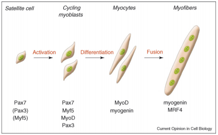
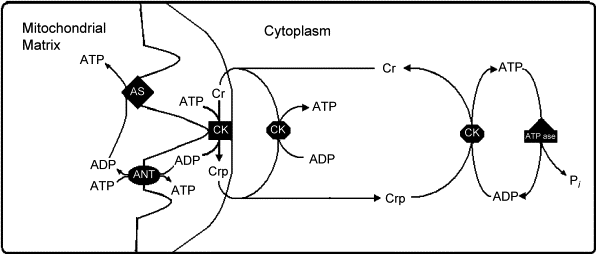
A critical side effect of doxorubicin treatment is myotoxicity. Treatment with doxorubicin has been found to downregulate myogenin and MyoD, two factors necessary for the production of muscle cells and the fusion of myoblasts (Yang, et al). This contributes to the loss of skeletal muscle and cardiac muscle.

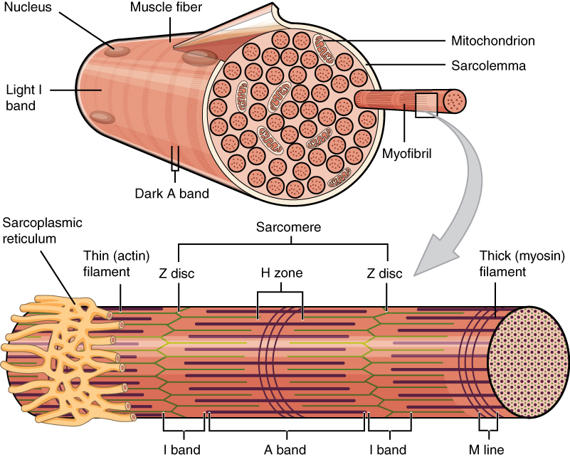
The current review will focus on the toxicity of doxorubicin toward skeletal muscle.

In normal muscle, the Myogenic Regulatory Factors function in specific order to activate satellite cells and form new muscle cells. All satellite cells produce Pax7, a factor essential for their maintenance (Le Grand, Fabien and Michael A Rudnicki). The mechanisms used to activate satellite cells remain a topic of study, but once activated, MyoD is produced within the cell (Le Grand, Fabien and Michael A Rudnicki). The activated satellite cells then leave the cell cycle (they lose the capability to perform mitosis) and will leave their initial location between the sarcolemma and the basal lamina (Le Grand, Fabien and Michael A Rudnicki). Myogenin and Mrf4 do not have the same function in satellite cell development, but myogenin production is necessary for the differentiation of “myotubes and fibers” (Le Grand, Fabien and Michael A Rudnicki). Myf5 is essential for the homeostasis and production of satellite cells.

MyoD is a helix-loop-helix transcription factor that is essential for satellite cell differentiation in skeletal muscle after such cells have been activated by phenomena such as mechanical stress (Le Grand, Fabien and Michael A Rudnicki). The Id2 gene is inducible by doxorubicin as a result of its 5’ flanking region and is expressed in proliferating tissues. Id2 gene expression, however, is downregulated during differentiation (Kurabayashi, et al.). Since muscle cells rely on the migration and differentiation of satellite cells, muscle will not generate sufficiently since doxorubicin is inducing Id2 to inhibit MyoD and therefore satellite cell differentiation (Kurabayashi, et al.).

Creatine, a compound widely used by bodybuilders to enhance performance and to increase muscle mass, may indeed be an effective treatment for myotoxicity side effects associated with doxorubicin chemotherapeutic treatment. Deldicque, et al. studied the effects of creatine supplementation on several myogenic factors in C2C12 cells. MyoD expression as well as myogenin expression were significantly enhanced in comparison with control C2C12 cells.

Creatine supplementation dramatically increases creatine as well as phosphocreatine content within the body. Consumption of 20g/day for 5-7 days has been found to increase creatine stores by about 20% and phosphocreatine stores by about 25% (Krieder). Creatine supplementation has been found to increase performance in a variety of activities. While around 70% of short-term supplementation studies show improvements in performance or muscle mass, around 90% of long-term supplementation studies show improvements ranging from 10-100% (Kreider). No clinically significant side effects were reported (Kreider).

Creatine is taken up by skeletal muscle cells by means of a sodium-ion dependent transporter protein which transports creatine against the concentration gradient when it is consumed orally (Willoughby & Rosene). As the concentration of intracellular creatine increases, so does the expression of the creatine transport protein as well as the dimeric muscle creatine kinase isozyme (Willoughby, Rosene). In turn, myosin heavy chain (MHC) and myosin expression are up-regulated. Since such changes in muscle cause the recruitment of Type I and II motor units for Type I and II muscle fibers, MyoD expression increases since it is associated with with Type IIb muscle fibers and the Type IIx MHC isoform (Willoughby & Rosene).

Creatine also functions in the regeneration of ATP particularly in muscle cells by means of the creatine-phosphate system. As ATP is produced by ATP synthase in the inner mitochondrial membrane, it exits to the intermembrane space in exchange for ADP via the Adenine Nucleotide Transporter (Brosnan, et al.). This ATP can then phosphorylate creatine at points where both mitochondrial membranes touch via creatine kinase, producing phosphocreatine and ADP (Brosnan, et al.). Creatine phosphate can then be used to regenerate ATP in the cytosol with creatine kinase and ADP (Brosnan, et al.).

In order to assess whether creatine supplementation is an effective treatment for doxorubicin-induced inhibition of muscle regeneration, the rat model should be used. After treatment with differing concentrations of creatine, rats can be analyzed for physical strength through grip strength tests, in which rats are suspended and their ability to maintain grip on a stationary object while being pulled is quantified, as well as other assessments. Homogenized rat tissue supernatant can be separated through gel electrophoresis and then analyzed for MyoD concentration as an indicator of muscle regeneration at the molecular level.

It is expected that creatine supplementation, as it upregulates MyoD expression and thereby contributes to muscle growth, will be an effective co-treatment with doxorubicin since it is likely to reverse the inhibitive effects of doxorubicin on muscle regeneration. If so, a statistically significant increase in MyoD concentration in rat tissue obtained from rats co-treated with doxorubicin and creatine should be noted from a control group treated with only doxorubicin. Rats treated with both creatine and doxorubicin should be able to exert more strength in grip tests and therefore measure greater force in such tests. Muscle obtained from rats treated with both creatine and doxorubicin should be able to exert more force than the control doxorubicin treatment.

Muscles of interest include the Extensor Digitorum Longus and the soleus muscle. These muscles were selected as they are representative of two major types of muscle, slow- and fast-twitch muscle, and can therefore be accurate models for the impacts creatine supplementation and doxorubicin treatment have on all parts of the body. The soleus is a slow-twitch muscle in the calf that extends the foot downward when the knee is bent, and the EDL is a superficial fast-twitch muscle in the leg that bends the foot upward at the ankle (The Editors of Encyclopedia Britannica 2011; 2018).